Case 2 - Buddy
According to the owner Buddy was playing with a ball, jumped up and landed funny. On physical examination pain and severe swelling is detected over right tarsus, specially on lateral aspect, non-weight bearing lameness.
Orthogonal xrays reveal a lateral malleolus fracture (Fig.1 and 2), stress views and palpation revealed stable joint with no affection of collateral ligaments but due to the fracture there is some degree of rotational instability. Surgical stabilization is strongly advised as Buddy is a 40 kg patient.
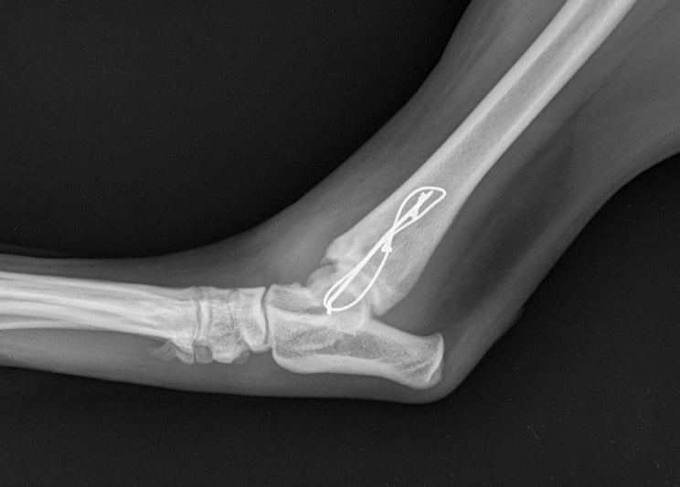
Lateral approach to the talocrural joint reveals severe oedema and bruising, 1.2mm IM pin placed in a distal to proximal fashion, 0.75mm cerclage wire used to create a figure-of-8 tension band. 1.4mm pin placed parallel to the talocrural joint from lateral to medial (Fig 3 and 4).
Buddy recovered well and in 4 weeks post op Xrays we could see that the fracture was healing well (Fig 5 and 6).
Fig. 2
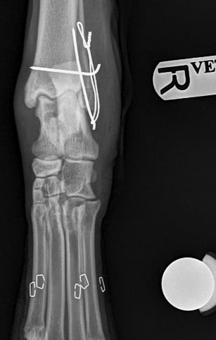

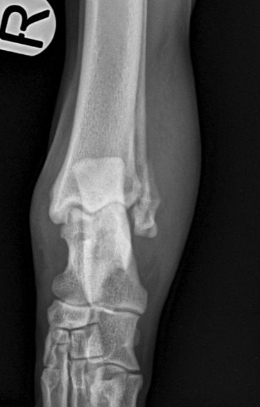

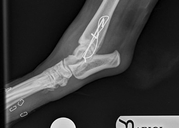

Lateral malleolus fractrure – Golden retriever, 2y, male
Fig. 1
Fig. 3
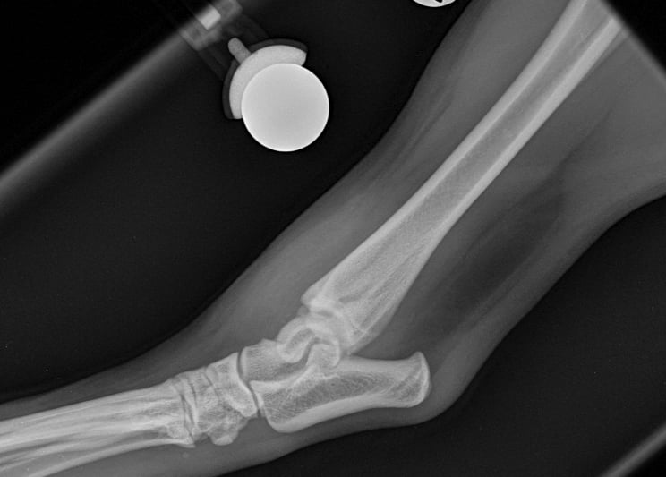

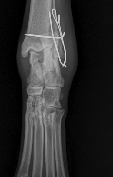

Fig. 6
Fig. 5
Fig. 4

Troya Surgery
Specialized mobile surgical services for veterinary practices.
Contact details
Email: troya.surgery@gmail.com
Telephone: 0771 625 1040
© 2024. All rights reserved.
WhatsApp: +44 771625 1040
