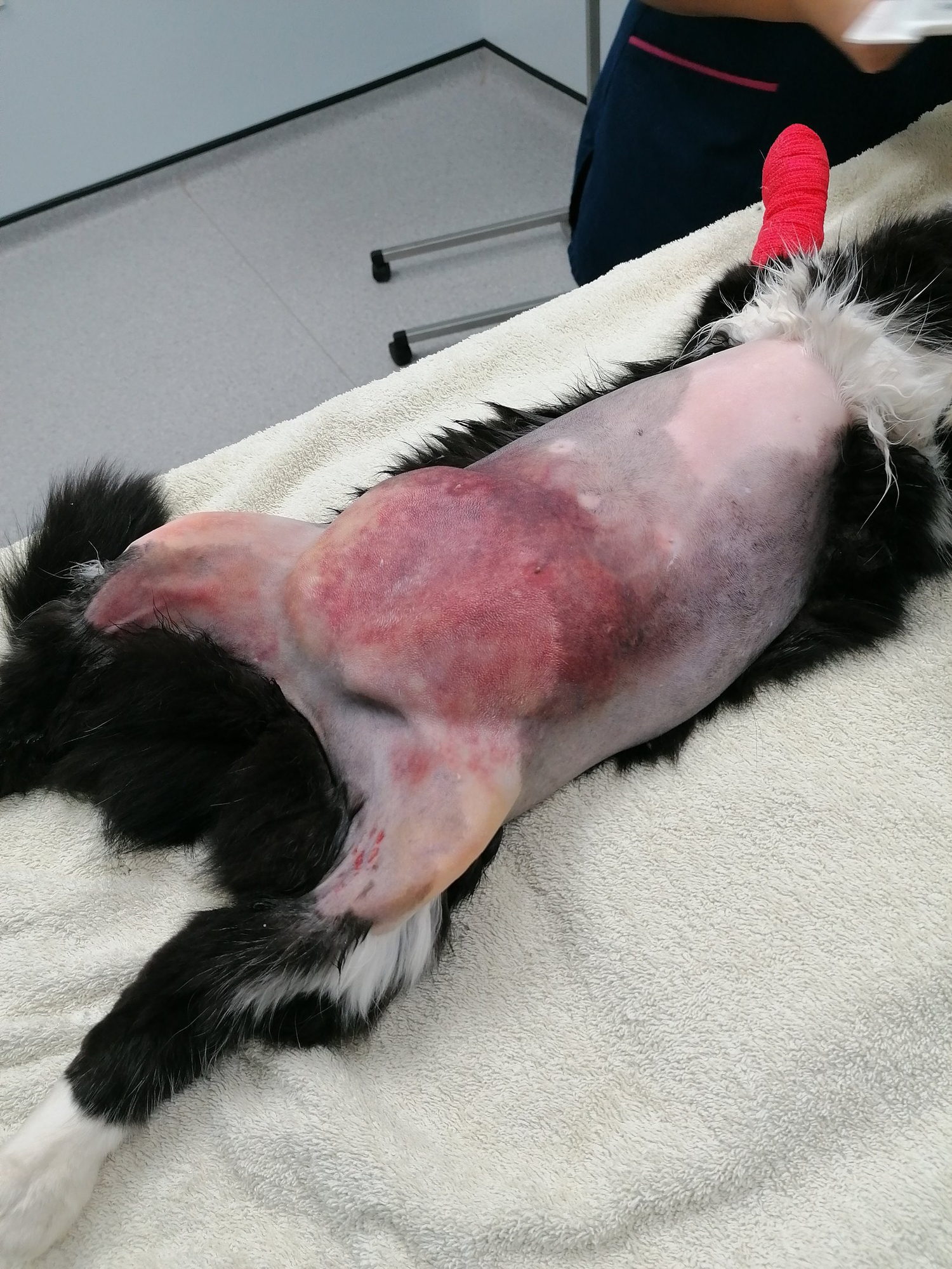
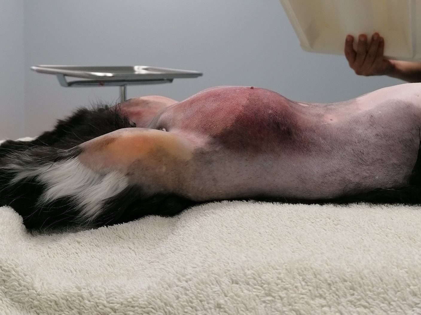
After being missing for five days, Jackson returned home with a noticeable lump on his abdomen. His owner observed that he was sleeping more than usual and appeared slightly lethargic. On examination, Jackson had pale pink mucous membranes and a large, pendulous abdomen.
Imaging & pre-op findings
Lateral X-rays confirmed the presence of a significant abdominal hernia. Upon clipping the area, the abdominal skin appeared extensively bruised.
Surgical findings & repair
A midline coeliotomy revealed severely swollen and bruised abdominal fat, with the abdominal musculature absent from 2 cm caudal to the umbilicus. The musculature was found retracted on both sides of the abdomen, and the prepubic abdominal tendon was torn and retracted cranially.
To repair the damage:
- Abdominal musculature mobilised caudally and reattached to the cranial edge of the pubic periosteum using 3.5-metric polypropylene
- Abdominal linea alba and both inguinal rings closed with 3-metric polydioxanone
- Jackson-Pratt drain placed to manage post-operative fluid
Outcome
Jackson remained in the hospital for two additional days and made an excellent recovery.