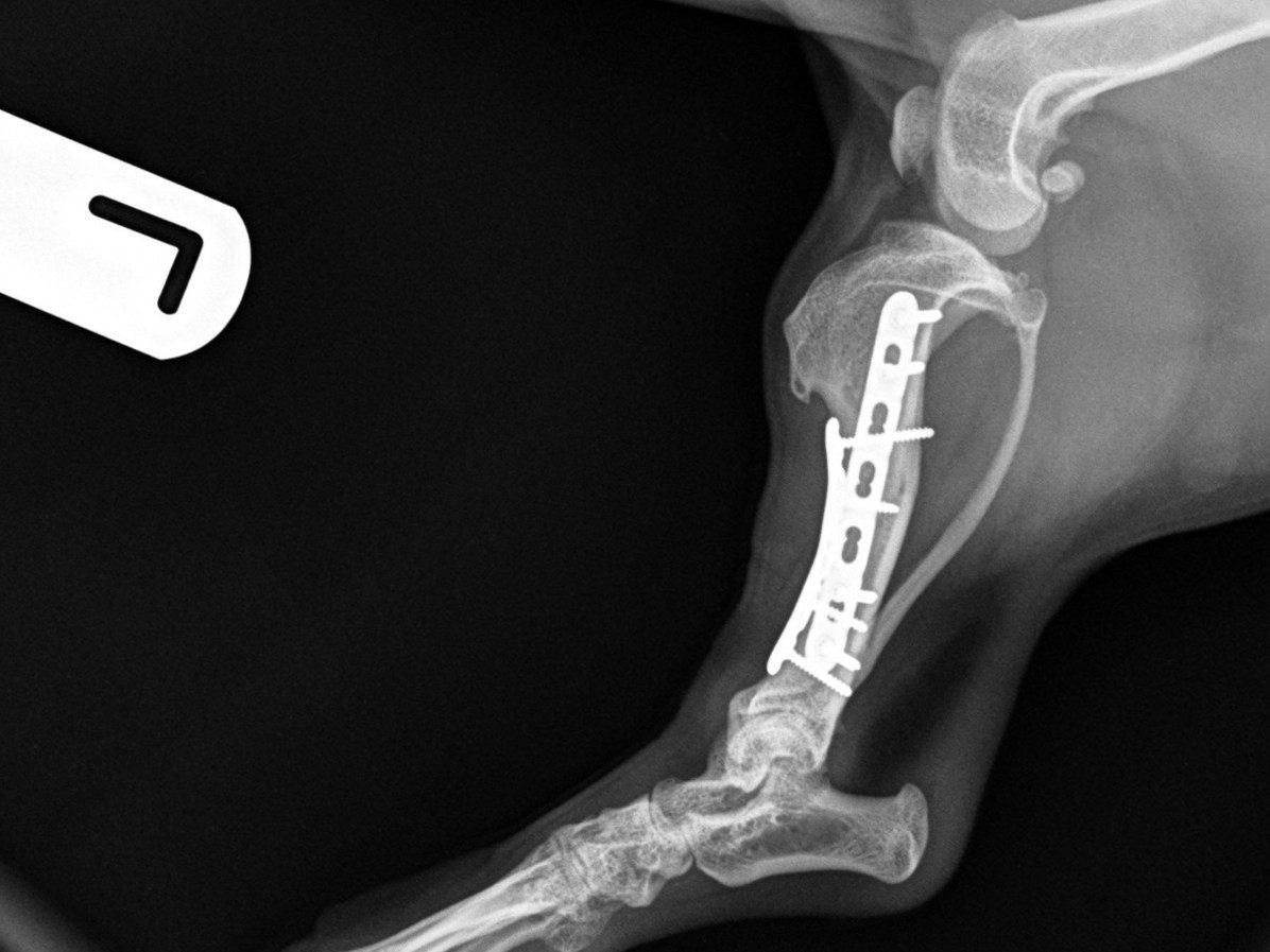
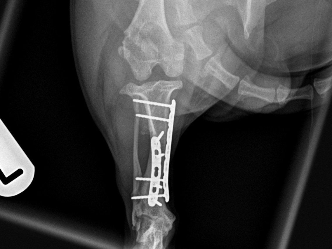
This small and sweet 5.4 kg female patient sustained a lameness after playing in the garden allotment. Orthogonal radiographs revealed a long-oblique spiral mid-diaphyseal fracture of the left tibia.
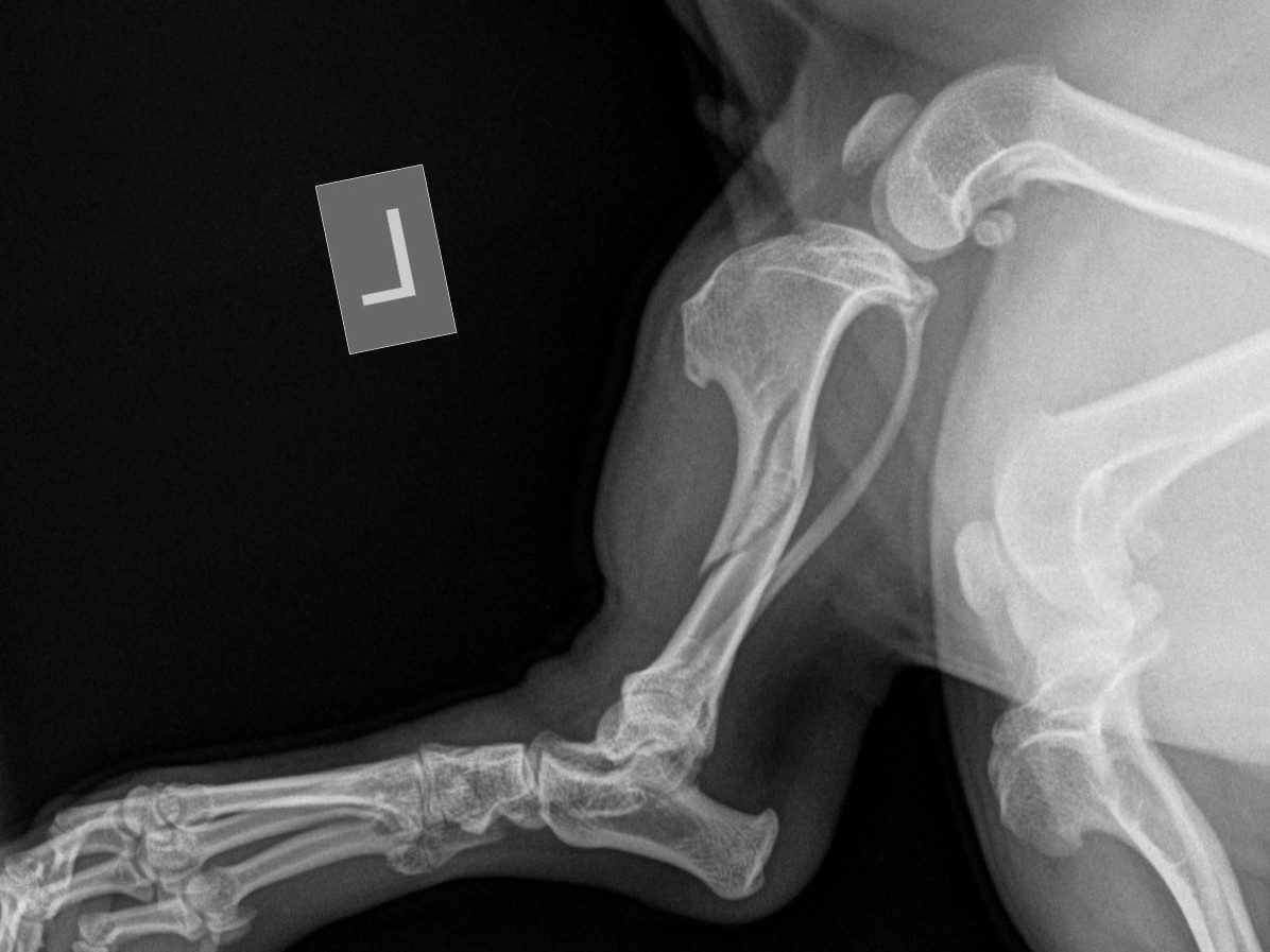
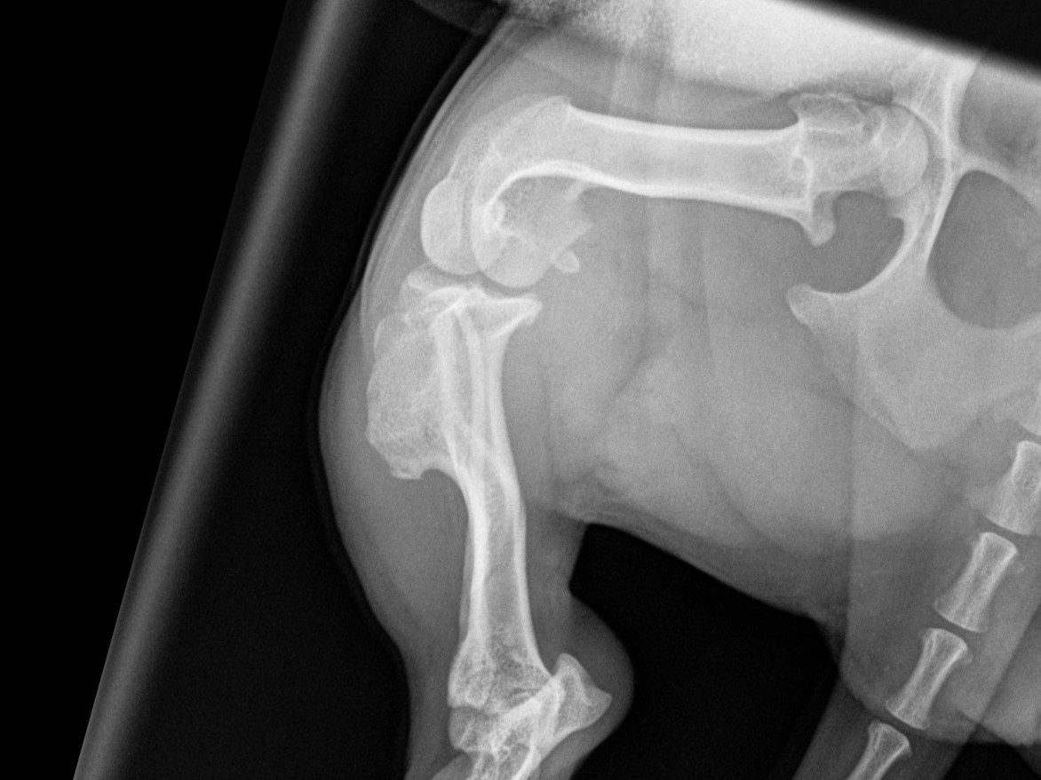
Surgical repair
A medial approach to the left tibial shaft was performed, with careful preservation of the saphenous vein throughout the procedure. The fracture site was identified and anatomically reduced using bone-holding forceps. Internal fixation was achieved using double plating in an orthogonal configuration, providing optimal mechanical stability.
Medial plate
A 1.5 mm 7-hole locking compression plate (LCP) applied in bridging fashion, secured with two bicortical locking screws in both the proximal and distal fracture fragments.
Cranial plate
A 1.5 mm 5-hole LCP also applied in bridging mode, with two locking screws in the proximal fragment (the most proximal bicortical and the distal monocortical due to proximity to the fracture line), and two bicortical locking screws in the distal fragment.
Closure
- Subcuticular layer: simple continuous pattern using 2-metric Monocryl
- Skin: intradermal closure with 2-metric Monocryl
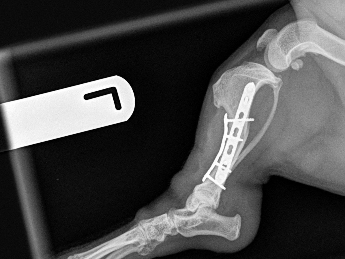
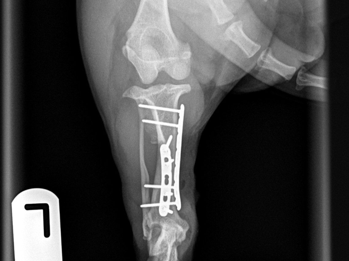
Six-week recheck
At the 6-week post-operative recheck, radiographs showed good progression of fracture healing with maintained alignment and implant integrity.