Luna presented with acute, severe lameness in her right pelvic limb following vigorous activity chasing a ball. On physical examination, there was localised pain and swelling at the right stifle joint. As the lameness failed to improve after a course of NSAIDs, radiographs were taken to investigate further.
Imaging
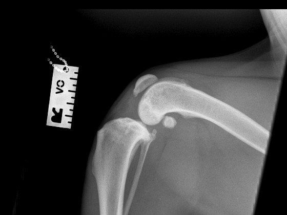
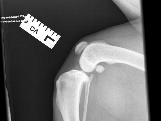
Orthogonal radiographs revealed findings consistent with cranial cruciate ligament disease. The lateral view of the affected limb shows joint effusion along with degenerative changes suggestive of osteoarthritis affecting the distal pole of the patella, proximal trochlear groove and fabellae. The contralateral (left) stifle appears normal and serves as a reference. A cranial drawer test was performed under sedation and yielded a positive result.
Surgical planning
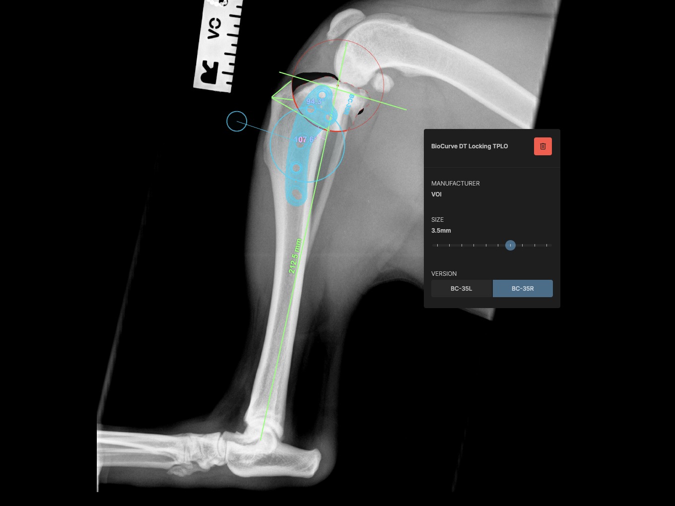
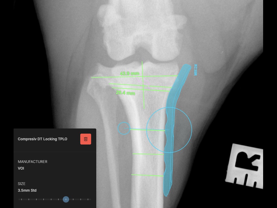
The surgical plan was developed using specialised veterinary orthopaedic planning software, allowing precise measurements and implant selection for optimal execution of the procedure.
Procedure
A sciatic nerve block with bupivacaine was administered as part of the analgesic protocol. Arthrotomy confirmed a complete rupture of the cranial cruciate ligament; remnants were debrided using a No. 11 scalpel blade. Both menisci were intact and stable upon probing. The caudal cruciate ligament was unremarkable. The joint was thoroughly lavaged and closed with interrupted cruciate sutures using 3-metric PDS.
Pre-operative measurements indicated a tibial plateau angle (TPA) of 25°. A TPLO saw blade size 24 was used, achieving a rotation of 8 mm. Pre-surgical measurements were: A = 11 mm, B = 13 mm, C = 35 mm.
A 3.5 mm TPLO locking plate was placed with two compression screws. A 2 mm anti-rotational pin was left in situ, as it did not interfere with the placement of the proximal fragment screws.
Closure
- Pes anserinus and fascia: simple continuous pattern with 3-metric PDS
- Subcuticular layer: simple continuous suture using 3-metric Monocryl
- Skin: intradermal pattern with 3-metric Monocryl
Post-operative imaging
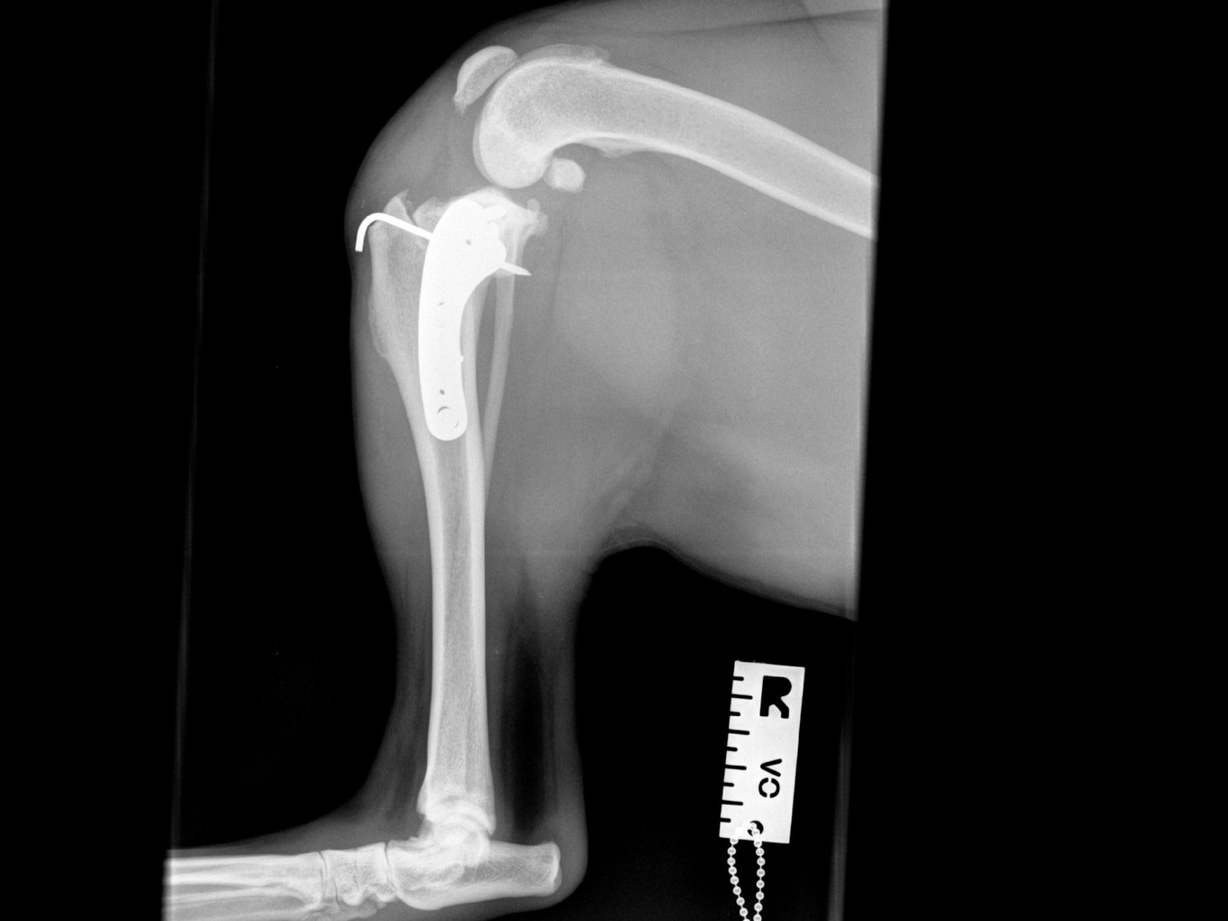
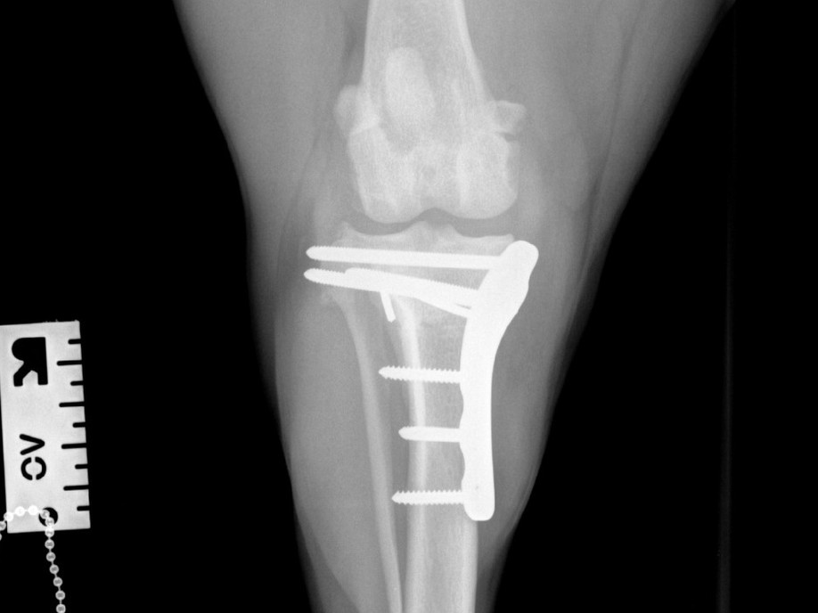
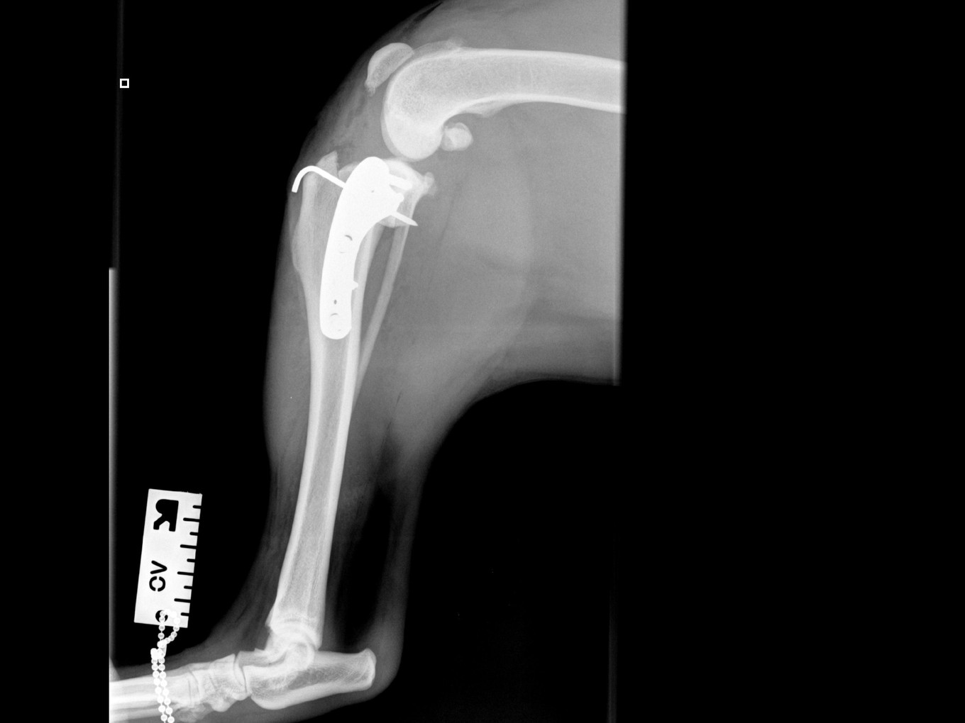
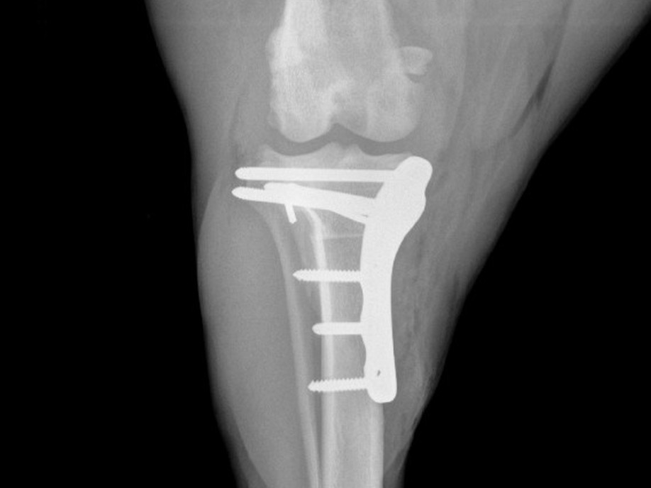
Post-operative radiographs showed that the proximal screw of the distal fragment is slightly longer than expected, but this is not anticipated to cause clinical issues. Implants are in good position, with correct compression and optimal alignment. Post-operative TPA measured at 4°.
Post-operative care plan
- Medications: NSAIDs for 14 days; Pardale for 5 days, then paracetamol to complete a 14-day course
- Monitoring: rechecks scheduled at 3 and 10 days post-op to assess wound healing and limb use
- Follow-up imaging: radiographic evaluation at 6 weeks
- Home care: follow printed discharge instructions; prevent wound licking — Buster collar if necessary
6-week follow-up
At the first post-operative recheck (day 3), Luna was nearly fully weight-bearing with only mild lameness. The surgical site showed excellent healing. Six-week post-op radiographs confirm good ossification of the osteotomy line and good position of implants.