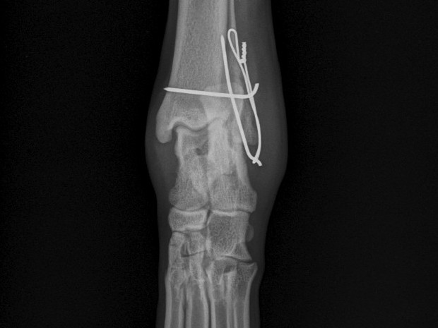
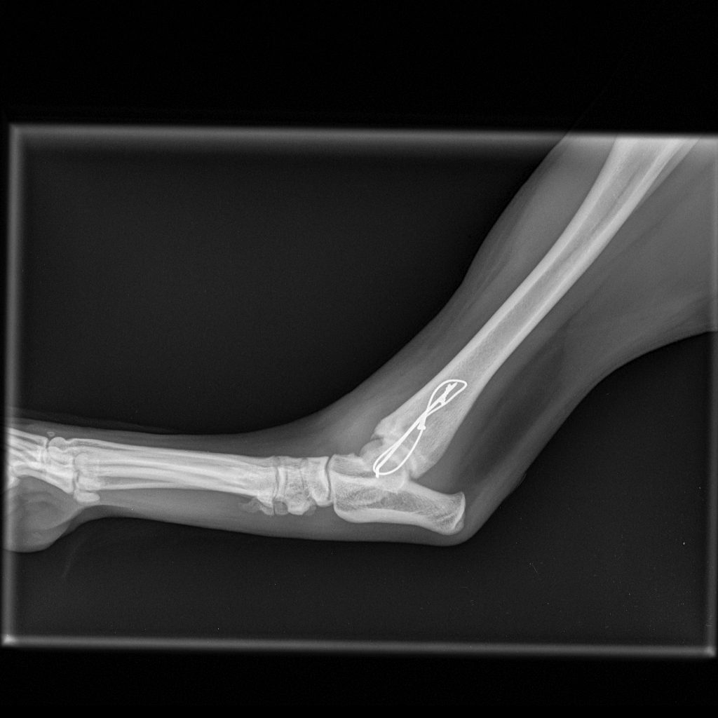
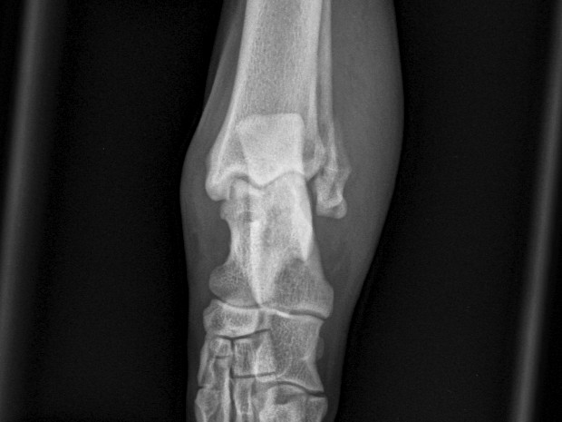
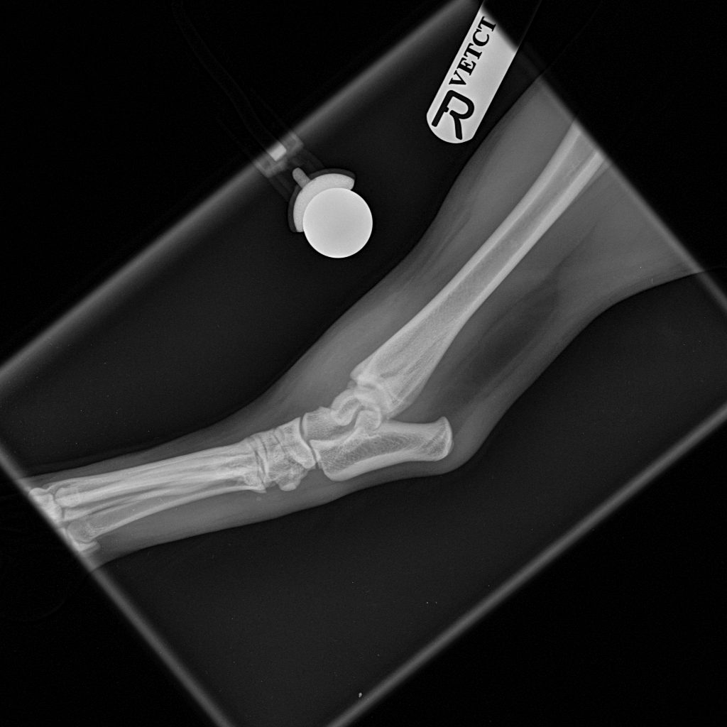
According to the owner, Buddy was playing with a ball, jumped up and landed funny. On physical examination, pain and severe swelling were detected over the right tarsus, especially on the lateral aspect, with non-weight-bearing lameness.
Imaging & assessment
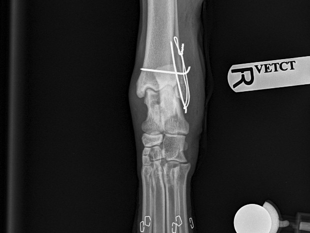
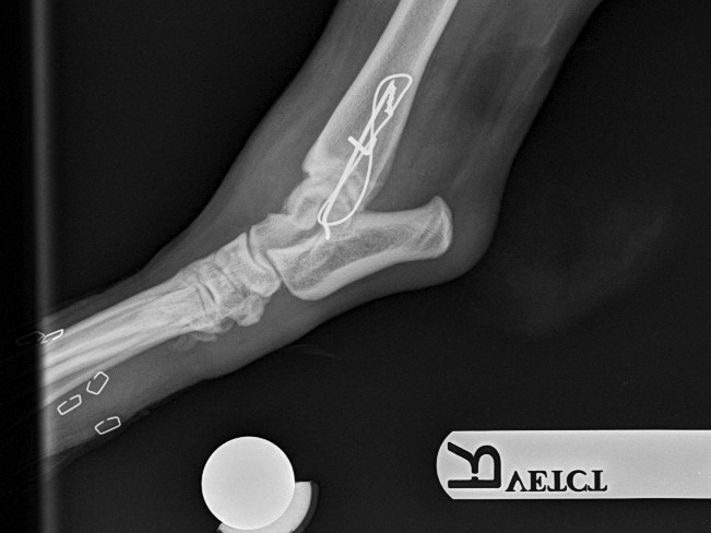
Orthogonal radiographs revealed a lateral malleolus fracture. Stress views and palpation confirmed a stable joint with no involvement of the collateral ligaments — but on manipulation, a mild to moderate degree of rotational instability was noted. Surgical stabilisation was strongly advised given Buddy’s 40 kg body weight.
Surgical repair
A lateral approach to the talocrural joint revealed severe oedema and bruising. Repair was achieved with:
- 1.2 mm IM pin placed in a distal-to-proximal fashion
- 0.75 mm cerclage wire used to create a figure-of-8 tension band
- 1.4 mm pin placed parallel to the talocrural joint, lateral to medial
Follow-up
Buddy recovered well, and at 4 weeks post-operative radiographs the fracture was healing well.