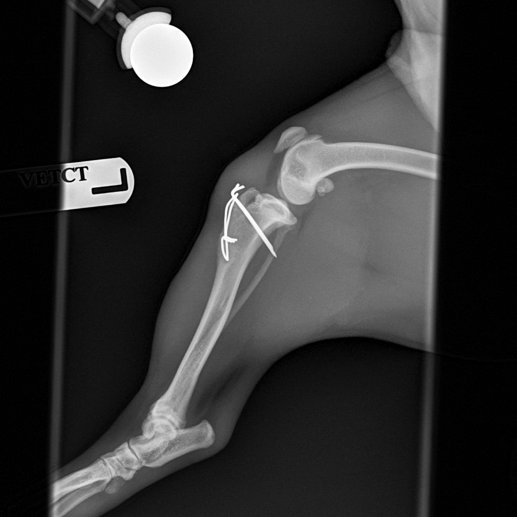
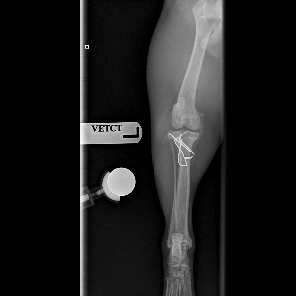
Ralph presented with non-weight-bearing lameness in his left pelvic limb following a jump over a baby gate. On examination, there was localised pain and inflammation over the tibial crest.
Imaging
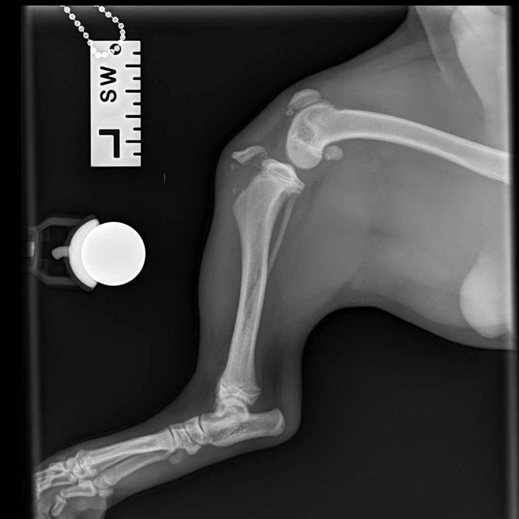
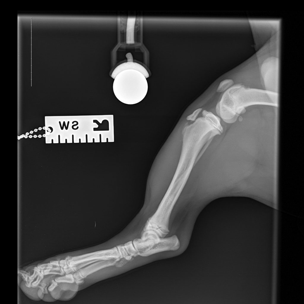
Lateral radiographs revealed a complete avulsion of the left tibial crest, along with a mild lesion at the distal pole of the patella. The contralateral right limb is shown for reference, demonstrating a normal appearance of the tibial crest growth plate.
Surgical repair
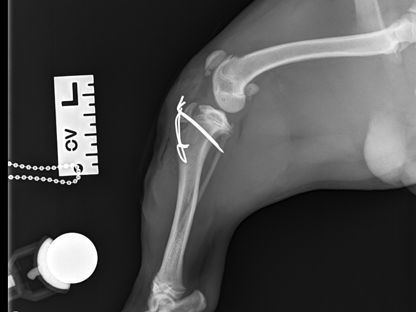
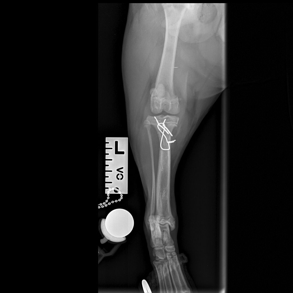
A medial approach to the proximal tibia was performed. The avulsed tibial tuberosity fragment was identified, anatomically repositioned and stabilised with two 1.2 mm Kirschner pins and a 1 mm tension-band wire.
Recovery
At the first post-operative recheck (3 days), Ralph was already weight-bearing with moderate lameness, and the surgical wound demonstrated excellent early healing. Six-week follow-up radiographs showed good ossification and healing of the fracture line, with progressive improvement also noted in the patellar avulsion lesion.